
An collaborator took saliva samples two minutes before and after an x-ray scan at his dentist’s. Coming back to the institute he compared the two drop samples under the microscope. The results show an evident difference in structure caused by the x-ray scan.
The objective of the test was to find out whether the impact of x-ray radiation on the human organism would substantially change saliva droplet structure, as seen under a microscope.
The test is was a random sampling. The test subject went in for a scheduled dentist appointment to have his back teeth x-rayed. The x-ray machine in question was rated at a 69 kV voltage, operating on a 15 mA current. Shortly before the x-ray scan the test subject took a sample of his saliva with a one-way syringe. The same was done two minutes after the scan.
On the way back to the institute the saliva was kept in the syringes for about two hours before being depositted on the glass slides. The photographs were taken as soon as the drops had dried in order to avoid any further changes in the saliva droplet structures.
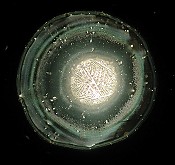
Normally 14 drops of a sample are deposited on the glass slides. The presented photographs show the 4 most characteristic samples.
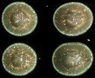
The droplet structures have distinctly changed. The bar structures are agglomerating towards the centre. The spaces between the border and centre of the droplets are largely empty forming concentric circles. This could be a direct consequence of tension or a defence mechanism of the living organism. Particularly interesting was the strongly increased production of saliva after the x-ray scan. Furthermore, this saliva was very viscid causing rather large dimensioned droplets.
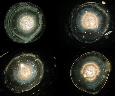
Two minutes after the scan the droplet structures have again completely changed. This was the moment the test subject felt emotionally at ease and calm. The saliva was again very fluid as was the case before the x-ray scan. Notice the slighlty visible concentric circles in between the large centre as well as the touch of bluish colouring to the intermediate area.
The tests show a clear impact of the x-ray scan on the saliva droplet structures.
One could further speculate that the droplet structures changed due to the influence of feelings, emotions and stress. At the moment of the test however, the tested subject was calm. Light emotion could be observed before the test due to the novelty of the situation, but it is unlikely that this had any major impact.
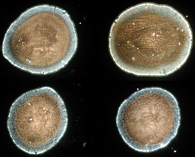
Surprisingly, the saliva largely regained its original structure just two minutes after the x-ray scan.
Looking at the last batch of photographs, the test subject recognized his very own “typical” saliva droplet structures.
You are kindly invited to give us your comments on the interpretation of the pictures and to communicate with us by dialog@weltimtropfen.de.
